

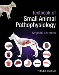
Textbook of Small Animal Pathophysiology
Beschreibung

The comprehensive overview of the essential discipline of pathophysiology in small animal disease
Textbook of Small Animal Pathophysiology provides a complete and accessible overview of disease mechanisms and pathophysiological consequences in the field of small animal medicine. The text focuses on the relationship between pathophysiology and disease of any etiology to aid better diagnostics and targeted therapy.
The text includes general pathophysiology of key organ systems such as cardiac, respiratory, gastrointestinal, neural, and more. Core information such as diagnostic methods and therapeutic options is presented in tabular form and clear full-color illustrations are featured throughout to aid in seamless reader comprehension.
Written by a highly qualified professional with significant firsthand experience in the field, Textbook of Small Animal Pathophysiology includes information on:
- Common pathological mechanisms, such as inflammation, sepsis and cytokine storm, fibrosis, degeneration, obesity, ischemia, hypoxia, cell death, apoptosis, necrosis, neoplasia, infection, ageing and pain
- General clinical manifestations, like abdominal enlargement, ataxia, constipation, cough, diarrhea, dyschezia, dysphagia, dyspnea, effusion, fatigue, fever, inappetence, jaundice, oedema, polyphagia, pruritus, regurgitation, seizure, shock, syncope, urinary disorders, vomitus, and weight loss
- Pathophysiology of organ systems cardiac system, hypertension, respiratory system, gastrointestinal system, hepatobiliary system, exocrine pancreas, urinary system, electrolyte system and acid-base balance, endocrine system, reproduction system, nerve system, joints, haematology and infectious diseases
Written to be a complete resource on the subject, Textbook of Small Animal Pathophysiology enables its readers to develop a clear understanding of how each disease works in order to improve decision making and patient outcomes. It is essential reading for veterinary students and a highly useful reference for small animal clinicians.
Weitere Details
Weitere Ausgaben
Andere Ausgaben
Person
The Author
Stephan Neumann Prof. Dr. med. vet. is Clinical Director of the Small Animal Clinic and apl. Prof. at the University of Göttingen, Germany.
Inhalt
Preface x
Abbreviations xii
Part I General Pathological Mechanisms 1
1 Inflammation 3
2 Sepsis and Cytokine Storm 14
3 Fibrosis 19
4 Degeneration 24
5 Obesity 27
6 Ischaemia, Hypoxia 32
7 Cell Death 36
8 Neoplasia 40
9 Infections 46
10 Ageing 53
11 Pain 58
12 Hypothalamus Function 61
Part II General Clinical Manifestations (Alphabetical) 65
13 Abdominal Enlargement 67
14 Ataxia 70
15 Constipation 74
16 Cough 78
17 Diarrhoea 81
18 Dyschezia 87
19 Dysphagia 90
20 Dyspnoea 93
21 Effusions 97
22 Fatigue 104
23 Fever 106
24 Inappetence 110
25 Jaundice 113
26 Oedema 116
27 Polyphagia 119
28 Pruritus 122
29 Regurgitation 124
30 Seizure 126
31 Shock 130
32 Syncope 133
33 Urinary Disorders 138
34 Vomiting 143
35 Weight Loss 147
Part III Pathophysiology of Organ Systems 151
36 Cardiac System, Hypertension 153
36.1 Physiological Functions and General Pathophysiology of Organ Insufficiency 153
36.2 Congenital Cardiac Diseases 161
36.3 Atrioventricular Valvular Diseases 167
36.4 Cardiomyopathy, Hypertrophic 172
36.5 Cardiomyopathy, Dilated 176
36.6 Heart Arrhythmia 181
36.7 Pericardial Disease 184
36.8 Hypertension 190
37 Respiratory System 195
37.1 Physiological Functions and General Pathophysiology of Organ Insufficiency 195
37.2 Brachiocephalic Syndrome 200
37.3 Acute Respiratory Distress Syndrome (ARDS) 203
37.4 Asthma (Feline Lower Respiratory Tract Disease) 207
37.5 Chronic Obstructive Bronchitis 210
37.6 Pneumonia 213
37.7 Idiopathic Lung Fibrosis 217
38 Gastrointestinal System 220
38.1 Physiological Functions and General Pathophysiology of Organ Insufficiency 220
38.2 Megaoesophagus 226
38.3 Gastritis/Ulceration 229
38.4 Gastric Dilatation-Volvulus Syndrome 233
38.5 Enteritis/Inflammatory Bowel Disease 237
38.6 Intestinal Lymphangiectasias 243
38.7 Intestinal Obstruction 249
39 Hepatobiliary System 253
39.1 Physiological Functions and General Pathophysiology of Organ Insufficiency 253
39.2 Acute Hepatic Failure 261
39.3 Chronic Hepatitis; Liver Fibrosis and Cirrhosis 266
39.4 Portosystemic Shunts/Hepatoencephalopathy 272
39.5 Feline Hepatic Lipidosis 277
39.6 Cholangiohepatitis 280
40 Exocrine Pancreas 284
40.1 Physiological Functions and General Pathophysiology of Organ Insufficiency 284
40.2 Acute Pancreatitis 287
40.3 Chronic Pancreatitis 292
40.4 Exocrine Pancreas Insufficiency 296
41 Urinary System 300
41.1 Physiological Functions of the Kidneys and General Pathophysiology of Organ Insufficiency 300
41.2 Glomerulonephritis/Nephrotic Syndrome 305
41.3 Tubular Disease/Fanconi Syndrome 310
41.4 Acute Renal Failure 313
41.5 Chronic Kidney Disease 321
41.6 Urinary Tract Infection 327
41.7 Urolithiasis 330
42 Electrolyte System and Acid-Base Balance 335
42.1 Acid-Base Balance 335
42.2 Hypo-/Hypernatraemia 340
42.3 Hypo-/Hyperkalaemia 344
43 Endocrine System 349
43.1 General Physiology 349
43.2 Pituitary Gland 352
43.2.1 Acromegaly 352
43.2.2 Pituitary Dwarfism 356
43.2.3 Diabetes Insipidus 359
43.3 Thyroid Gland 362
43.3.1 Physiology of the Thyroid Gland 362
43.3.2 Hyperthyroidism 365
43.3.3 Hypothyroidism 369
43.4 Parathyroid Gland 374
43.5 Endocrine Pancreas 379
43.5.1 Physiology of Endocrine Pancreas 379
43.5.2 Diabetes Mellitus 382
43.5.3 Diabetic Ketoacidosis 387
43.5.4 Insulinoma 390
43.6 Adrenal Gland 393
43.6.1 Physiology of the Adrenal Gland 393
43.6.2 Hyperadrenocorticism 397
43.6.3 Hypoadrenocorticism 402
43.6.4 Hyperaldosteronism 405
43.6.5 Pheochromocytoma 409
44 Reproduction System 413
44.1 Pyometra 413
44.2 Prostate Diseases 417
45 Nerve System 420
45.1 Brain Tumours 420
45.2 Idiopathic Epilepsy 423
45.3 Intervertebral Disc Disease 426
45.4 Peripheral Neuropathy 429
46 Joints 431
46.1 Arthritis 431
46.2 Osteoarthritis 434
47 Haematology 437
47.1 Physiological Functions of Red Blood Cells 437
47.2 Erythrocytosis 441
47.3 Anaemia 444
47.4 Coagulation Disorders 450
47.5 Hypercoagulability 454
48 Infectious Diseases 457
48.1 Feline 457
48.1.1 Feline Leukaemia Virus Infection 457
48.1.2 Feline Infectious Peritonitis 461
48.1.3 Feline Immunodeficiency Virus Infection 466
48.2 Canine 471
48.2.1 Distemper 471
48.2.2 Canine Parvovirus 475
48.3 Bacterial Diseases 480
48.3.1 Anaplasmosis 480
48.3.2 Borreliosis 484
48.3.3 Ehrlichiosis 488
48.3.4 Leptospirosis 491
48.4 Protozoal Infections 495
48.4.1 Babesiosis 495
48.4.2 Giardiasis 499
48.4.3 Leishmaniasis 503
49 Common Immune-mediated Diseases 507
49.1 Autoimmunity 507
49.2 Lupus Erythematosus 510
49.3 Myasthenia Gravis 514
50 Common Clinical Biochemical Parameters (Alphabetical) 517
50.1 Alanine Aminotransferase (ALT) 517
50.2 Albumin 518
50.3 Alkaline Phosphatase (ALP) 520
50.4 Ammonia 522
50.5 Aspartate Aminotransferase (AST) 523
50.6 Bile Acids 524
50.7 Bilirubin 525
50.8 Calcium 526
50.9 Cholesterol 528
50.10 Creatinine 529
50.11 Creatine Kinase 530
50.12 Glucose 531
50.13 Glutamate Dehydrogenase (GLDH) 532
50.14 Lipase 533
50.15 Phosphorus 534
50.16 Potassium 535
50.17 Sodium 537
50.18 Total Protein 539
50.19 Urea 541
Index 542
1
Inflammation
Definition
Inflammation is by definition a defensive reaction of the body with the aim of eliminating the trigger of the inflammation and repairing the damage caused, either by restoring the tissue of origin (restitutio) or by a replacement tissue, the scar (reparatio).
Trigger
Inflammation is triggered by any stimuli, called 'noxae', that overcome the organism's defence-compensation mechanisms. By definition, a noxious agent is a substance or event that causes damage to a biological organism. The noxious agent can be divided according to its origin into internal and external, and according to its structure into physical, chemical or biological triggers (Table 1.1).
Defence Cascade of the Body
Numerous defence mechanisms exist to protect an organism. These are particularly effective at the 'contact surfaces' between the organism and the environment. There, the penetration of the noxious agent is prevented by mechanical as well as biological defence mechanisms.
The skin prevents the penetration of the noxious agent under physiological conditions due to its structural design. For this purpose, the skin consists of different layers with different functions (Figure 1.1).
The outer layer of the skin, the epidermis, is made up of the layers - stratum corneum, lucidum, granulosum, spinosum and basale from the outside inwards. The main cell type in the epidermis is the keratinocyte. This differentiates in the stratum basale from epidermal stem cells. In the stratum spinosum, the cells begin to remodel with an increase in volume and a change in shape and width. In the further course, keratohyalin grains are formed in the stratum granulosum, and further remodelling processes take place. The cells become flattened, the nucleus is lost, shrinkage occurs due to fluid loss, and finally cornification takes place. Eventually, no more keratinocytes can be detected in the stratum corneum. Keratinocytes become corneocytes. The cornification process builds up a mechanical protection for the skin. In addition, penetration of a noxious substance is ensured by a close connection between the keratinocytes through tight-junctions. The tight-junctions consist of connections of transmembrane proteins, such as claudin and occludin. Intracellularly, these proteins are connected to the cytoskeleton. The tight-junctions connect the cells into a bandage that forms a barrier to the paracellular penetration of a noxious agent.
In addition, defence cells such as Langerhans cells are localised in the epidermis.
These are tissue macrophages that are capable of phagocytosis but also differentiate into antigen-presenting cells after contact with an antigenically active noxious agent, which can initiate an immune response.
Finally, the extracellular matrix in the epidermis forms a molecular association that can prevent the penetration of a noxious agent. The molecules of the extracellular matrix include, for example, keratins and collagens as structural proteins and ceramides, which are lipids composed of a sphingosine molecule and fatty acids that provide protection against hydrophilic noxae (Mitamura et al. 2021) (Table 1.2).
Table 1.1 Common causes of inflammation in dogs and cats.
Physical Chemical Biological Infectious agents Heat Environmental toxins Viruses Cold Endogenous toxins Tumours Bacteria Degenerations Fungi ParasitesFigure 1.1 Structure of the outer skin and development of keratinocytes.
Table 1.2 Components of the epidermal extracellular matrix with function.
Molecule Function Keratin Intermediate filament, structural element of keratinocytes Collagen Tight protein of the ECM, adhesion molecule Elastin Elastic protein of the ECM Fillagrin Structural protein Ceramide Hydrophobic protectionThe mucous membranes of the body form the inner boundary layer between the organism and the environment. The microscopic structure of the mucous membranes already reflects defence competences. This includes the contact of the mucosal cells through tight-junctions. These form a tight connection between the cell membrane of neighbouring cells through proteins such as occludin. This prevents the paracellular penetration of extracorporeal noxae.
Another superficial defence mechanism is the synthesis of mucins. Mucins are glycoproteins that are synthesised by goblet cells and form a protective layer several micrometres thick on the mucosa. In the process, defence functions of the mucins develop due to their gel-like structure, which enables mechanical protection of the underlying mucosal cells. Chemically, for example, bicarbonate residues of the mucins can bind and inactivate acids, and biologically, mucins can prevent bacteria from invading by binding them.
Cells localised in the mucosa (Paneth cells) secrete lysozyme or defensins to inactivate germs. The latter are differentiated into a-, ß- and ?-defensins based on their molecular structure (Lehrer and Ganz 2002).
The effects of defensins are antimicrobial and immunomodulatory. The former effect is based on their positive molecular charge, which enables a charge-dependent interaction with the negatively charged bacterial cell wall, especially the lipopolysaccharides (Scott and Hancock 2000). The binding of the defensins results in pore formation. The consequence is a depolarisation of the bacterial cell membrane and thus a breakdown of the membrane potential and lysis of the cell (Scott and Hancock 2000; Sahl et al. 2005).
In addition, defensins show immunomodulatory functions. Defensins have a chemotactic effect on dendritic cells and memory T cells, and thus represent a link between innate and adaptive immune responses. In addition, some defensins act chemotactically on monocytes and macrophages, and in some cases induce mast cell activation and degranulation. As a result, histamine and prostaglandins are released, which promote the migration of neutrophilic granulocytes. Degranulation of the recruited neutrophilic granulocytes in turn releases defensins again, resulting in a positive feedback loop (Yang et al. 2002).
The secretion of lysozyme is another defence mechanism against bacterial invasion of the mucous membranes. Lysozyme is an enzyme that cleaves murein. Murein is a peptidoglycan and a component of the bacterial cell wall. Due to the hydrolytic cleavage of murein, the bacterial cell wall loses its selective permeability and rupture of the bacterial cell wall occurs due to increased water influx.
Another defence mechanism in the mucosa is the mucosa-associated lymphoid system located in the lamina propria, which is called 'gut associated lymphoid tissue' (GALT) in the gastrointestinal tract and bronchial associated lymphoid tissue (BALT) in the respiratory tract. This system includes numerous cells of the non-specific and specific defence response, such as macrophages and lymphocytes (Figure 1.2).
Figure 1.2 Building up the defence system of the mucous membrane.
Player of the Inflammation
The inflammatory process is maintained by some so-called 'inflammatory cells'. These combine some properties that predispose them to fulfil the definitional task of inflammation. This states that inflammation is a reaction of the body that serves to eliminate a noxious agent and its consequences.
The following cells are involved in the inflammatory process:
Neutrophilic granulocytes originate from the leukocyte pool of the bone marrow and are distributed throughout the blood. The residence time in the blood is a few hours (6-12 hours). Subsequently, the neutrophilic granulocytes leave the blood capillaries under the influence of chemoattractive substances. These are released as part of the local inflammatory process. The process of neutrophilic granulocyte emigration from the blood vessels takes place via adhesion and transmigration. Integrin-mediated, the neutrophilic granulocytes adhere to the surfaces of the endothelial cells. In the process, the cells change their shape from roundish to an amoeboid cell shape. The cells can now migrate trans- and paracellularly through the vascular endothelial layer.
In the area of inflammation, the neutrophilic granulocytes have different functions. The ability to phagocytose allows the cells to take up and lyse the noxious agent (e.g. bacteria). For this process, the noxious agent is taken up by membrane invagination and the resulting vesicle combines with the granular structures of the granulocyte. In...