

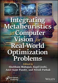
Integrating Metaheuristics in Computer Vision for Real-World Optimization Problems
Beschreibung

A comprehensive book providing high-quality research addressing challenges in theoretical and application aspects of soft computing and machine learning in image processing and computer vision.
Researchers are working to create new algorithms that combine the methods provided by CI approaches to solve the problems of image processing and computer vision such as image size, noise, illumination, and security. The 19 chapters in this book examine computational intelligence (CI) approaches as alternative solutions for automatic computer vision and image processing systems in a wide range of applications, using machine learning and soft computing.
Applications highlighted in the book include:
- diagnostic and therapeutic techniques for ischemic stroke, object detection, tracking face detection and recognition;
- computational-based strategies for drug repositioning and improving performance with feature selection, extraction, and learning;
- methods capable of retrieving photometric and geometric transformed images;
- concepts of trading the cryptocurrency market based on smart price action strategies; comparative evaluation and prediction of exoplanets using machine learning methods; the risk of using failure rate with the help of MTTF and MTBF to calculate reliability; a detailed description of various techniques using edge detection algorithms;
- machine learning in smart houses; the strengths and limitations of swarm intelligence and computation; how to use bidirectional LSTM for heart arrhythmia detection;
- a comprehensive study of content-based image-retrieval techniques for feature extraction;
- machine learning approaches to understanding angiogenesis;
- handwritten image enhancement based on neutroscopic-fuzzy.
Audience
The book has been designed for researchers, engineers, graduate, and post-graduate students wanting to learn more about the theoretical and application aspects of soft computing and machine learning in image processing and computer vision.
Weitere Details
Weitere Ausgaben
Andere Ausgaben
Personen
Shubham Mahajan, PhD, is an assistant professor in the School of Engineering at Ajeekya D Y Patil University, Pune, Maharashtra, India. He has eight Indian, one Australian, and one German patent to his credit in artificial intelligence and image processing. He has authored/co-authored more than 50 publications including peer-reviewed journals and conferences. His main research interests include image processing, video compression, image segmentation, fuzzy entropy, nature-inspired computing methods with applications in optimization, data mining, machine learning, robotics, and optical communication.
Kapil Joshi, PhD, is an assistant professor in the Computer Science & Engineering Department, Uttaranchal Institute of Technology in Dehradun, India. His doctorate was on image quality enhancement using fusion techniques. He has 8 years of academic experience and has published patents, research papers, and two books. In 2021, he was awarded the 'Best Young Researcher' Award in Global Education and Corporate Leadership received by Life Way Tech India Pvt. Ltd.
Amit Kant Pandit, PhD, is an associate professor in the School of Electronics & Communication Engineering Shri Mata Vaishno Devi University, India. He has authored/co-authored more than 60 publications including peer-reviewed journals and conferences. He has two Indian and one Australian patent to his credit in artificial intelligence and image processing. His main research interests are image processing, video compression, image segmentation, fuzzy entropy, and nature-inspired computing methods with applications in optimization.
Nitish Pathak, PhD, is an associate professor in the Department of Information Technology, Bhagwan Parshuram Institute of Technology, New Delhi, India. He has 17 years of engineering education experience and has published more than 80 journal articles, in peer-reviewed journals as well as book chapters, patents, and conference papers. His research areas include intelligent computing techniques, empirical software engineering, and artificial intelligence.
Inhalt
Preface xv
1 Advancement in Diagnostic and Therapeutic Techniques for Ischemic Stroke 1
Mukul Jain, Divya Patil, Shubham Gupta and Shubham Mahajan
1.1 Introduction 2
1.2 Diagnostic Tools of Ischemic Stroke 4
1.3 Artificial Intelligence-Based Diagnostic Tools 7
1.4 Blood-Based Protein Biomarker for Stroke 8
1.5 Markers for Endothelial Damage 8
1.6 Markers of Brain Injury 9
1.7 Therapeutic Advances in Ischemic Stroke 9
1.8 Nanoparticles 11
1.9 Conclusion 13
2 Object Detection and Tracking Face Detection and Recognition 25
Varsha K. Patil, Pawan Nawade, Rudra Nagarkar and Paresh Kadale
2.1 Introduction 25
2.2 Motivation 30
2.3 The Basics of Computer Vision 31
2.4 Face Detection 34
2.5 Facial Expression 38
2.6 Object Detection 41
2.7 Face Detection and Identification in Practical Situations 44
2.8 Future Direction in Object Detection and Tracking 47
2.9 Conclusion 52
3 Printing Organs with 3D Technology 55
Shaik Aminabee
3.1 Introduction 55
3.2 Bioprinting in Three Dimensions (3D) 56
3.3 3D Printing Types 57
3.4 Applications for 3D Printing in Cells 60
3.5 New Developments 65
3.6 Progress in India 66
3.7 Limitation 67
3.8 A Future Point of View 67
3.9 Conclusion 68
4 Comparative Evaluation of Machine Learning Algorithms for Bank Fraud Detection 71
Kiran Jot Singh, Divneet Singh Kapoor, Kunal Ranjan Singh, Chirag Kalucha, Gatik Alagh, Khushal Thakur and Anshul Sharma
4.1 Introduction 71
4.2 Proposed Framework 73
4.3 Results 74
4.4 Concluding Remarks and Future Scope 77
5 An Overview of Computational-Based Strategies for Drug Repositioning 81
Shalu Verma, Nidhi Nainwal, Alka Singh, Gauree Kukreti and Kiran Dobhal
5.1 Introduction 81
5.2 Drug Repositioning 82
5.3 Challenges and Opportunities for Drug Repurposing 93
5.4 Conclusion 94
6 Improving Performance With Feature Selection, Extraction, and Learning 99
Varsha K. Patil, Vrinda Shinde, Ritika Singh and Vipul Singh
6.1 Introduction 99
6.2 Feature Selection 100
6.3 Feature Extraction 110
6.4 Feature Learning 115
6.5 Future Research and Development 123
6.6 Future Scope 124
6.7 Conclusion 125
7 Fusion of Phase and Local Features for CBIR 129
Pooja Sharma
7.1 Introduction 129
7.2 Overview of the Proposed System 132
7.3 Proposed Hybrid-Shape Descriptors 132
7.4 Similarity Measurement 137
7.5 Experimental Study and Performance Evaluation 139
7.6 Conclusions 147
8 Trading Bot for Cryptocurrency Market Based on Smart Price Action Strategies 151
Divneet Singh Kapoor, Kiran Jot Singh, Anshoom Jain, Rhythm Chauhan, Khushal Thakur and Anshul Sharma
8.1 Introduction 151
8.2 Background 154
8.3 Proposed Framework 156
8.4 Results 158
8.5 Conclusion and Future Scope 161
9 Comparative Evaluation and Prediction of Exoplanets Using Machine Learning Methods 163
Divneet Singh Kapoor, Kiran Jot Singh, Ashirvad Singh, Benarji Mulakala, Karan Singh, Prashant, Ramanjeet Singh and Shubham Mahajan
9.1 Introduction 164
9.2 Background 167
9.3 Proposed Framework 169
9.4 Results 171
9.5 Conclusion and Future Scope 182
10 The Risk of Using Failure Rate With the Help of MTTF and MTBF to Calculate Reliability 185
Harpreet Kaur and Shiv Kumar Sharma
10.1 Introduction 185
10.2 Failure 186
10.3 Conclusion 191
11 A Detailed Description on Various Techniques of Edge Detection Algorithms 193
Pritha A. and G. Fathima
11.1 Introduction 193
11.2 Edge Detection Techniques 194
11.3 Experimental Results 203
11.4 Comparative Results 203
11.5 Conclusion 203
11.6 Future Work 204
12 Advancement of ML in Smart House 207
Gokula Udhayan V., K. Mahaeshwari and N. Vinoth Kumar
12.1 Objective 207
12.2 Introduction 207
12.3 Smart House System With IoT 208
12.4 Future Scope 223
12.5 Conclusion 223
13 Multi-Robot Navigation: A Biologically Inspired Framework 225
Imran Mir and Faiza Gul
13.1 Introduction 225
13.2 Optimization Algorithms 226
13.3 Algorithms and Self-Organization 236
13.4 Future Research Directions 238
13.5 Conclusion 239
14 Bidirectional LSTM for Heart Arrhythmia Detection 243
Nikhil M. Agrawal, H. D. Bhanu Cheitanya, Abhishek Kumar Rai and Shubham Mahajan
14.1 Introduction 243
14.2 About the Dataset 245
14.3 Flow of the Model 246
14.4 Results 248
14.5 Conclusion 248
15 Study on Content-Based Image Retrieval 253
Thanga Subha Devi M., R. Suji Pramila and Tibbie Pon Symon
15.1 Introduction 254
15.2 Related Works 256
15.3 Extraction of Features 261
15.4 User Interactions for CBIR System 266
15.5 Conclusions 269
16 Machine Learning and Angiogenesis in Cancer 273
Dharambir Kashyap, Riya Sharma, Neelam Goel and Vivek Kumar Garg
16.1 Introduction 273
16.2 History of Angiogenesis Discovery 274
16.3 Overview of Angiogenesis 274
16.4 Angiogenesis in Carcinogenesis 275
16.5 Molecular Mechanisms of Angiogenesis Formation 276
16.6 Angiogenesis as a Target in Cancer Therapy 276
16.7 Machine Learning Approaches in Angiogenesis 277
16.8 Conclusion 278
17 Handwritten Image Enhancement Based on Neutroscopic-Fuzzy and K-Mean Clustering 283
Jaspreet Kaur, Divya Gupta, Simarjeet Kaur and Amrinder Singh
17.1 Introduction 284
17.2 Application of Image Processing 286
17.3 Enhancement of Handwritten Document 287
17.4 Clustering Techniques 288
17.5 Performance Parameters 290
17.6 Results and Discussion 293
17.7 Conclusion 295
18 A Texture Classification System Based on an Adaptive Histogram Equalized Shearlet Transform 299
K. Gopalakrishnan, V. Karthikeyan and P.T. Vanathi
18.1 Introduction 299
18.2 Literature Survey 303
18.3 Materials and Methods 305
18.4 Proposed Methodology 309
18.5 Result and Discussion 311
18.6 Conclusion 320
19 A Thyroid Nodule Detection Using L1-Norm Inception Deep Neural Network 323
Saranya G.
19.1 Introduction 323
19.2 Related Work 324
19.3 Methodology 325
19.4 Results and Discussion 329
19.5 Conclusion 336
References 337
Index 339
1
Advancement in Diagnostic and Therapeutic Techniques for Ischemic Stroke
Mukul Jain1,2, Divya Patil2, Shubham Gupta3 and Shubham Mahajan4,5,6*
1Cell & Developmental Biology Lab, Research and Development Cell, Parul University, Vadodara, Gujarat, India
2Department of Life Sciences, Parul Institute of Applied Sciences, Parul University, Vadodara, Gujarat, India
3Department of Paramedical and Health Sciences, Faculty of Medicine, Parul University, Vadodara, Gujarat, India
4School of Engineering, Ajeenkya D Y Patil University, Pune, Maharashtra, (iNurture Education Solutions Pvt. Ltd., Bangalore), Pune, Maharashtra, India
5University Center for Research & Development (UCRD), Chandigarh University, Mohali, India
6Hourani Center for Applied Scientific Research, AI-Ahilyaya Amman University, Amman, Jordan
Abstract
Ischemic stroke is a cerebrovascular disease that is typically brought on by a disruption in the blood flow to the brain; it accounts for more than 80% of all strokes. 1.8 million neurons are thought to die per minute. The burden of stroke in people younger than 65 years has increased over the last decades, it has increased worldwide by 25% among adults aged 20 to 64 years. According to reports, both men and women can experience stroke symptoms, in this case women are more prone to have nontraditional symptoms of stroke like dizziness, lack of consciousness, slurred speech. In comparison to Western Europe, America, Australia, and similar to Eastern Europe, Asia has a higher stroke fatality rate. Acute management of stroke requires fast and efficient screening, imaging and modality. Better clinical outcomes can be attained through early detection and treatment of stroke. Stroke imaging techniques are computed tomography (CT) scans and magnetic resonance imaging (MRI). MRI is more preferred because it gives detailed and exact result of damage. Antithrombotic and neuroprotective therapies are the mainstays of conventional medicine, although their use is still constrained due to their poor safety. Utilizing nanomedicines is an additional option since they can boost therapeutic benefit and adverse effect reduction, achieve effective medicine collection at the target site, and improve pharmacokinetic behavior of pharmaceuticals in vivo. We have comprehensively described ischemic stroke, its epidemiology, advanced artificial intelligence-based diagnostic tools and its treatment therapies.
Keywords: Ischemic stroke, computed tomography, MRI, artificial intelligence, diagnostic tools, PET, EEG, nanomedicine
1.1 Introduction
Stroke is a fatal and disabling illness that affects more than 15 million individuals annually worldwide. It is an ageing disease that primarily affects persons over the age of 65 years [1, 2]. In the US, ischemic strokes, which are caused by a decrease in brain blood circulation, account for 87% of all cases [2]. According to a report released by WHO in 2016, strokes are the second most frequent cause of impairment in the population today worldwide (Figure 1.1). With the passing of each decade during the previous 40 years, it has been revealed that the number of stroke cases multiplied [11]. Both the incidence and prognosis of stroke are influenced by gender; nevertheless, overall, women have a higher stroke prevalence than men do due to aging-related increases in stroke risk and longer average lifespans for women [3]. Basic stroke classifications include hemorrhagic, transient ischemic attack, and ischemic strokes (Figure 1.2). Nearly 80% of strokes are ischemic, while various populations experience hemorrhagic and ischemic strokes more frequently in varied proportions [4]. The following categories-etiologic subtypes-have been used to further categorize ischemic strokes: cardioembolic, atherosclerosis, lacunar, other particular causes (dissections, vasculitis, specific hereditary illnesses, etc.), and strokes with no known etiology [5]. Prior to the main stroke episode, individuals always have ministrokes, commonly referred to as transient ischemic attacks (TIA). The majority of published research relies on MRI and CT scan pictures to categorize strokes, which is a costly method of early stroke detection. CT and MRI are employed in the majority of investigations that identify strokes. These days, noninvasive methods are becoming more and more common. One such method is the electroencephalograph (EEG), which is also a cheap or free method. The following issues have impeded the advancement of novel therapeutics for ischemic stroke and numerous other illnesses of the central nervous system (CNS): blood-brain barrier (BBB) prevents most CNS drugs from reaching the brain parenchyma effectively, which makes it difficult to treat or save areas of the brain that haven't yet been completely affected; (2) many drugs have poor stability or toxicity after systemic and/or oral administration; (3) the physiopathology of the disease is not well understood; and (4) it is challenging to translate positive results from preclinical studies to the clinic [6]. One of the key problems in the treatment of stroke is the blood brain barrier. In order to transport enough medicine to the brain tissue, liposomes must be able to cross the BBB and it is ideal for them to stay in the bloodstream for a long time [7]. New stroke diagnostic methods have been created thanks to nanotechnology and artificial nanomaterials. Identifying the biochemical and pathophysiological causes of stroke may be made easier with the help of this technology [8]. Due to their distinct fluorescence characteristics, which enable high spatiotemporal accuracy at biologically relevant concentrations, optical CNT-based biosensors exhibit considerable potential [9, 10]. With the development of nanotechnology, it is now possible to carry drugs to the brain and, more precisely, the area that is ischemia, more efficiently [8]. The FDA has only approved one therapeutic drug-recombinant tissue plasminogen activator (tPA)-for the treatment of ischemic stroke, and its use is constrained by its limited therapeutic window, quick drug elimination, and potential for hemorrhagic transformation [138]. There is no particular cure in medical science to deal with strokes; therefore, early identification is the key to deal with strokes. The early identification can inhibit the disabilities, loss of deaths, and other brain-related severe problems [12].
Figure 1.1 The data represent country wise occurrence of stroke and mortality rate in male and female across the world. (a) Incidence of stroke worldwide, in which china has higher percentage rate that is 9% and lower rate in Saudi Arabia that is 1%. (b) Prevalence of ischemic stroke across the world covering 28 countries, the higher incidence of ischemic stroke is 10% and low rate is 2%. (c) The percentage of mortality rate of stroke in male across the globe is 4%, high mortality rate is reported in Madagascar that is 9%. (d) The percentage of mortality rate in female across the world, high mortality is noted in Madgascar, Afghanistan, and Indonesia that is 9%.
Figure 1.2 It demonstrates the different types of stroke occurrence and how it causes in the brain and disturbs the brain function.
1.2 Diagnostic Tools of Ischemic Stroke
1.2.1 Preimaging
Patients with acute neurological conditions require quick neuroimaging. American Stroke Association recommendations recommend that the only prior inquiry is an actual capillary blood glucose, which is retrieved from paramedics. What is an intravenous cannula for contrast or perfusion imaging is frequently necessary sequences, enabling the simultaneous collection of a blood panel. This often includes a coagulation screen, a renal function test, and an infection test if the patient uses anticoagulants. Though many departments of radiology demand a current renal function prior to providing contrast [10].
1.2.2 Imaging
1.2.2.1 Computed Tomography Scan
Neuroimaging in the setting of a hyperacute severe stroke is still primarily based on CT. A noncontrast CT scan of head is rapid, delicate, and cost-effective excluding cerebral bleeding, which is typically sufficient to make judgments on thrombolysis. Due to widespread availability and significantly shorter turnaround times, CT perfusion investigations are extraeffective in the accident scenario to detect and identify stroke patients who can profit from thrombolytic therapy and vascular blood flow [134, 135] CT perfusion sequences can evaluate a variety of brain perfusion is frequently performed using artificial intelligence, like MIstar (Apollo Medical Imaging Technology) or iSchemaView's Rapid Processing of Perfusion and Diffusion (RAPID) CT perfusion. The contrast bolus-tracking approach known as CT perfusion can be used with almost any multidetector CT scanner that is now in use in emergency rooms all around the world [131, 132]. A crucial factor in determining the dangers and possible benefits of reperfusion treatments, ideal CT perfusion values and thresholds that characterize irreversible infarction have not received enough attention in research [133]. These technologies make interpretation easier by ensuring the use of validated thresholds and enhancing interobserver reproducibility [10]. The sensitivity of the CT signals is between 40% and 60% within the first 3 hours following the beginning of symptoms,...