

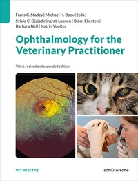
Ophthalmology for the Veterinary Practitioner
Beschreibung

This fully revised and expanded edition illustrates common ophthalmic diseases in dogs, cats, horses, birds, and farm animals. It covers clinical examination procedures, typical symptoms, breed-specific predispositions as well as medications and treatment options - both surgical and non-surgical - relevant to veterinary practitioners.
Structured to mirror the steps of a clinical examination , the book serves as a reliable and indispensable handbook both for the veterinary ophthalmology novice and the general practitioner.
The book highlights diseases that can be challenging to diagnose without specialized experience or specific instruments. Ophthalmic emergencies and rare presentations are also described to complete the range of possible ophthalmic disorders - making differential diagnosis easier for the veterinary practitioner.
Weitere Details
Weitere Ausgaben
Nachauflagen
Andere Ausgaben
Personen
Inhalt
- Front Cover
- Copyright
- Table of Contents
- Body
- Editors and authors
- Figure sources and credits
- Abbreviations
- 1 Introduction
- 2 Clinical and differential diagnostic procedures
- 2.1 Description of the patient
- 2.2 Patient history
- 2.3 Animal handling, equipment, and instruments
- 2.3.1 Restraint and sedation
- 2.3.2 Materials and instruments
- 2.4 Assessment of visual function
- 2.5 Examination of the eye and its adnexa
- 2.5.1 Head, skull, and orbital area
- 2.5.2 Tear film and tear production
- 2.5.3 Ocular discharge
- 2.5.4 Eyelids (palpebrae)
- 2.5.5 Conjunctiva
- 2.5.6 Globe (bulbus oculi)
- 2.5.7 Sclera
- 2.5.8 Cornea
- 2.5.9 Anterior and posterior chambers
- 2.5.10 Pupil and iris
- 2.5.11 Lens
- 2.5.12 Vitreous
- 2.5.13 Fundus
- 2.5.13.1 Morphology of the fundus
- 2.5.14 Additional and specific examinations
- 2.5.14.1 Chromatic pupillary light responses
- 2.5.14.2 Electroretinography (ERG) and visual evoked potentials (VEPs)
- 2.5.14.3 Diagnostic imaging techniques: radiographic, ultrasonographic, CT, and MRI examinations
- 2.5.14.4 Confocal scanning laser ophthalmoscopy (cSLO) and OCT
- 2.6 Differential diagnosis
- 2.6.1 Introduction
- 2.6.2 Epiphora without distinct blepharospasm
- 2.6.3 Blepharospastic/painful eye with sufficient or excessive tear production
- 2.6.4 Protrusion of the nictitating membrane with enophthalmos
- 2.6.5 Exophthalmos
- 2.6.6 The "red" eye
- 2.6.7 The "blue-white" cornea
- 2.6.8 The "pigmented" eye
- 2.6.9 The "blind" eye
- 3 Therapeutics, diagnostics, and surgical principles for ophthalmic diseases
- 3.1 Introduction
- 3.1.1 The conjunctival sac
- 3.1.2 Subconjunctival
- 3.1.3 Retrobulbar
- 3.1.4 Intraocular
- 3.1.5 General rules
- 3.2 Ocular therapeutic and diagnostic agents
- 3.2.1 Vasoconstrictors
- 3.2.2 Antiallergic agents (generally replaced by glucocorticosteroids today)
- 3.2.3 Antiglaucoma agents
- 3.2.3.1 Moderating production of aqueous: carbonic anhydrase inhibitors
- 3.2.3.2 Osmotic agents
- 3.2.3.3 ß-blocking agents
- 3.2.3.4 Prostaglandin analogues
- 3.2.3.5 Sympathomimetics
- 3.2.3.6 Miotics. Improving drainage of aqueous
- 3.2.4 Mydriatics
- 3.2.5 Antimicrobial agents
- 3.2.5.1 "Initial choice" antimicrobials/disinfectants
- 3.2.5.2 "Second choice" antibacterials
- 3.2.5.3 "Specific choice" antibacterials
- 3.2.5.4 Antimycotics
- 3.2.5.5 Virostatic drugs: DNA-synthesis inhibitors
- 3.2.6 Glucocorticosteroids
- 3.2.7 Administration
- 3.2.7.1 Topical, into the conjunctival sac
- 3.2.7.2 Subconjunctival
- 3.2.7.3 Oral
- 3.2.8 Non-steroidal anti-inflammatory drugs (NSAIDs)
- 3.2.9 Topical anesthetics
- 3.2.10 Vitamins, epithelializing agents, and neutral agents
- 3.2.11 Eye wash solutions (Collyria)
- 3.2.12 Other "drugs" for ocular use
- 3.2.12.1 Diagnostic agents
- 3.2.12.2 Mucolytic and discharge-dissolving agents
- 3.2.12.3 Antihypertensive agents (in secondary retinopathy)
- 3.2.12.4 Other drugs for ophthalmic use (additional reading, Herring 2021)
- 3.2.13 Radiation
- 3.2.14 Protective devices
- 3.3 Surgical options
- 3.3.1 Anesthesia
- 3.3.2 Preparation of the surgical field
- 3.3.3 Positioning on the operating table
- 3.3.4 Draping
- 3.3.5 Magnifying equipment
- 3.3.6 Surgical equipment
- 3.3.7 Suture material
- 3.3.8 Suturing
- 3.3.9 Hemostasis
- 3.3.10 Cryosurgery
- 3.3.11 Laser techniques
- 4 Ocular emergencies
- 4.1 Introduction
- 4.2 Luxation or proptosis of the globe
- 4.3 Chemical burns
- 4.4 Blunt trauma
- 4.4.1 Orbital fractures
- 4.4.2 Contusion of the globe
- 4.4.2.1 Suffusion (hyposphagma)
- 4.4.2.2 Traumatic corneal edema
- 4.4.2.3 Hyphema
- 4.4.2.4 Blunt trauma with deeper penetration
- 4.5 Penetrating or perforating trauma
- 4.5.1 Lid lacerations and conjunctival sac wounds
- 4.5.1.1 Lacerations of the lid margin including the lacrimal canaliculus
- 4.5.1.2 Lacerations with loss of tissue
- 4.5.2 Conjunctival lacerations
- 4.5.3 Corneal lacerations
- 4.5.3.1 General rules of treatment
- 4.5.3.2 Non-perforating corneal wounds
- 4.5.3.3 Perforating corneal defects
- 5 Orbit and globe
- 5.1 Introduction
- 5.2 Clinical presentation of orbital diseases
- 5.2.1 Enophthalmos
- 5.2.2 Exophthalmos
- 5.2.3 Luxation or proptosis of the globe or bulbus
- 5.2.4 Strabismus
- 5.3 Causes of orbital diseases
- 5.3.1 Atrophy of orbital/periorbital tissue
- 5.3.2 Sympathetic denervation/Horner's syndrome
- 5.3.3 Masticatory muscle myositis
- 5.3.4 Orbital muscle myositis
- 5.3.5 Other retrobulbar processes
- 5.3.6 Orbital fat prolapse/Prolapse of the Harderian gland
- 5.4 Clinical presentation and causes of bulbar diseases
- 5.4.1 Anophthalmia, cyclopia, microphthalmia
- 5.4.2 Phthisis bulbi
- 5.4.3 Macrophthalmos
- 5.4.4 Buphthalmos/hydrophthalmos
- 5.4.5 Endophthalmitis, panophthalmitis
- 5.4.6 Nystagmus
- 5.5 Surgery of globe and orbit
- 5.5.1 Enucleation of the globe or bulbus
- 5.5.1.1 Lateral subconjunctival approach
- 5.5.1.2 Transpalpebral approach
- 5.5.2 Evisceratio bulbi
- 5.5.3 Exenteratio orbitae
- 5.5.4 Orbitotomy
- 6 Lacrimal secretory and drainage systems
- 6.1 Introduction
- 6.2 Diseases of the lacrimal secretory system
- 6.2.1 Quantitative tear film deficiencies Keratoconjunctivitis sicca (KCS)
- 6.2.2 Qualitative tear film deficiencies
- 6.2.3 (Sialo)dacryoadenitis
- 6.2.4 Cysts and neoplasms
- 6.2.5 Prolapse of the nictitating membrane gland and the Harderian gland
- 6.3 Diseases of the nasolacrimal drainage system
- 6.3.1 Atresia, micropunctum, stenosis of the lacrimal puncta
- 6.3.2 Atresia and secondary closure of the canaliculi
- 6.3.3 Dacryocystitis
- 6.3.4 Lacerations and other reasons for obstruction
- 6.3.5 Cysts and neoplasms
- 7 Eyelids
- 7.1 Introduction
- 7.2 Ankyloblepharon
- 7.3 Palpebral aplasia
- 7.4 Dermoids/dysplasia of the lid
- 7.5 Distichiasis
- 7.6 Entropion
- 7.6.1 Entropion in sheep and horses
- 7.7 Ectropion and/or macroblepharon (oversized palpebral fissure)
- 7.7.1 Shortening of the lower palpebral conjunctiva
- 7.7.1.1 Basting the ventral palpebral conjunctiva
- 7.7.1.2 V-Y method
- 7.7.2 Simple wedge resection
- 7.7.3 Kuhnt-Szymanowski method
- 7.7.4 Kuhnt-Szymanowski method, Blaskovics modification (additional reading, Blaskovics 1959)
- 7.7.5 Z-plasty/free transplants
- 7.7.6 Total fissure shortening methods
- 7.8 Blepharophimosis
- 7.9 Trichiasis
- 7.9.1 Nasal fold trichiasis, medial entropion, macroblepharon, associated with shallow orbits
- 7.9.1.1 Removal of nasal folds
- 7.9.1.2 Medial canthoplasty
- 7.9.2 Upper eyelid trichiasis/entropion
- 7.9.3 Caruncle trichiasis and trichiasis in other locations
- 7.10 Injuries
- 7.11 Ptosis
- 7.12 Lagophthalmos
- 7.13 Blepharitis
- 7.13.1 Non-specific blepharitis
- 7.13.2 Chronic blepharitis
- 7.13.3 Specific blepharitis
- 7.13.3.1 Chalazion/hordeolum
- 7.13.3.2 Blepharitis adenomatosa (meibomianitis)
- 7.13.3.3 Juxtapalpebral defects/granulomatous changes
- 7.13.3.4 Eosinophilic granuloma
- 7.13.3.5 Blepharitis in birds
- 7.13.3.6 Blepharitis in horses
- 7.14 Neoplasia of the eyelids
- 7.14.1 Sarcoids in horses
- 8 Conjunctiva and nictitating membrane
- 8.1 Introduction
- 8.2 Non-pigmented margin of the nictitating membrane
- 8.3 Dermoid
- 8.4 Ectopic cilia
- 8.5 Protrusion of the nictitating membrane
- 8.6 Cysts
- 8.7 Eversion/inversion of the nictitating membrane
- 8.8 Prolapse of the gland of the nictitating membrane ("cherry eye")
- 8.9 Subconjunctival hemorrhages
- 8.10 Conjunctivitis
- 8.10.1 Serous/catarrhal conjunctivitis
- 8.10.2 Purulent conjunctivitis
- 8.10.3 Follicular conjunctivitis
- 8.10.4 Plasmacellular conjunctivitis of the nictitating membrane
- 8.10.5 Papillary/nodular/granulomatous conjunctivitis
- 8.10.6 Neonatal conjunctivitis (ophthalmia neonatorum)
- 8.10.7 Allergic conjunctivitis
- 8.10.8 Lipogranulomatous conjunctivitis
- 8.10.9 Infectious bovine/ovine keratoconjunctivitis (pink eye)
- 8.11 Eosinophilic granuloma
- 8.12 Symblepharon
- 8.13 Conjunctival stricture in the rabbit
- 8.14 Neoplasia of the conjunctiva
- 9 Cornea and sclera
- 9.1 Corneal diseases and disorders
- 9.1.1 Introduction
- 9.1.2 Clinical signs
- 9.1.3 Location of lesions and causes
- 9.1.4 Regeneration
- 9.1.5 Delayed epithelialization
- 9.2 Congenital corneal disorders
- 9.2.1 Microcornea
- 9.2.2 Persistent pupillary membrane (PPM)
- 9.2.3 Dermoid
- 9.3 Corneal trauma
- 9.4 Inflammatory corneal disease/keratitis
- 9.4.1 Superficial keratitis (non-ulcerative)
- 9.4.1.1 Chronic superficial keratitis/pannus (Überreiter's disease)
- 9.4.1.2 Eosinophilic keratitis
- 9.4.1.3 Pigmentary keratopathy/pigmentary keratitis
- 9.4.2 Deep or interstitial keratitis/keratitis profunda (non-ulcerative)
- 9.5 Corneal defects, erosions, and ulcers
- 9.5.1 Corneal defects
- 9.5.1.1 Spontaneous chronic corneal epithelial defect
- 9.5.1.2 Punctate keratitis
- 9.5.1.3 Ulcerative corneal disorders
- 9.5.1.4 Deep corneal ulcers
- 9.6 Corneal sequestrum
- 9.7 Corneal perforation
- 9.8 Infectious corneal diseases
- 9.8.1 (Herpetic) keratitis
- 9.8.2 Infectious bovine/ovine keratoconjunctivitis
- 9.8.3 Mycotic corneal infections
- 9.8.4 Corneal abscess (horse)
- 9.9 Degenerative/dystrophic corneal disorders
- 9.9.1 Degenerative corneal disorders
- 9.9.2 Corneal dystrophies
- 9.9.2.1 Epithelial/stromal dystrophy
- 9.9.2.2 Corneal macular dystrophy
- 9.9.2.3 Endothelial dystrophy, endothelial decompensation
- 9.9.3 Corneal inclusion cysts
- 9.10 Scleral diseases
- 9.10.1 (Epi)scleritis
- 9.10.2 Scleral neoplasia
- 10 Intraocular pressure and glaucoma
- 10.1 Introduction
- 10.2 Glaucoma
- 10.2.1 Classifications of glaucoma
- 10.2.1.1 Etiology
- 10.2.1.2 Stage of disease
- 10.2.1.3 Classification of primary glaucoma based on the appearance of the iridocorneal angle
- 10.2.2 Clinical aspects of glaucoma
- 10.2.2.1 Clinical signs of glaucoma
- 10.2.3 Primary glaucoma
- 10.2.3.1 Acute or insidious onset: Primary open-angle glaucoma (POAG)
- 10.2.4 Treatment
- 10.2.4.1 Medical treatment
- 10.2.4.2 Surgical treatment and photocoagulation
- 10.2.5 Secondary glaucoma
- 10.2.5.1 Secondary glaucoma associated with the lens or vitreous
- 10.2.5.2 Secondary glaucoma associated with uveal changes
- 10.2.5.3 Secondary glaucoma associated with hyperpigmentation, ocular (uveal) melanosis and melanocytic glaucoma
- 10.2.5.4 Secondary glaucoma associated with trauma
- 10.2.5.5 Secondary glaucoma associated with intraocular neoplasia
- 10.2.5.6 Secondary glaucoma associated with medication
- 10.2.5.7 Secondary glaucoma associated with intraocular surgery
- 11 Uvea
- 11.1 Introduction
- 11.1.1 Iris
- 11.1.2 Ciliary body
- 11.1.3 Choroid
- 11.2 Persistent (epi)pupillary membrane
- 11.3 Iris hypoplasia
- 11.4 Acorea
- 11.5 Heterochromia of the iris
- 11.6 Blue iris/white coat
- 11.6.1 Color dilution and ocular abnormalities
- 11.6.2 Coat, iris color and deafness
- 11.6.3 Partial oculocutaneous albinism
- 11.7 Color changes in the iris
- 11.8 Uveal cysts
- 11.9 Hyphema
- 11.9.1 Dysplastic vascular abnormalities
- 11.9.2 Trauma
- 11.9.3 Vascular wall injury
- 11.9.4 Systemic arterial hypertension
- 11.9.5 Coagulation disorders
- 11.9.6 Anterior uveitis
- 11.9.7 Neoplasms
- 11.10 Anterior uveitis (posterior uveitis)
- 11.10.1 Traumatic uveitis
- 11.10.2 Metabolic uveitis
- 11.10.3 Infection-related uveitis
- 11.10.3.1 Viral
- 11.10.3.2 Bacterial
- 11.10.3.3 Ehrlichia and rickettsia
- 11.10.3.4 Mycotic
- 11.10.3.5 Algae
- 11.10.3.6 Protozoal
- 11.10.3.7 Nematodes
- 11.10.4 Immune-mediated uveitis
- 11.10.4.1 Lens-induced uveitis
- 11.10.4.2 Uveodermatologic syndrome (UDS)
- 11.10.4.3 Lupus erythematosus (LE)
- 11.10.5 Idiopathic uveitis
- 11.10.6 Pseudo-uveitis caused by neoplasia
- 11.10.7 Equine recurrent uveitis
- 11.10.8 Heterochromic iridocyclitis and keratitis (HIK) in the horse
- 11.10.9 Anterior uveitis in the rabbit
- 11.11 Iris atrophy
- 11.12 Dysautonomia or pupillary dilatation syndrome (Key-Gaskell syndrome)
- 11.13 Horner's syndrome
- 11.14 Other pupillary abnormalities
- 11.15 Neoplasia
- 11.16 Posterior uvea
- 12 Lens and vitreous
- 12.1 Introduction
- 12.1.1 Ontogenesis
- 12.1.2 Anatomy and physiology of the lens
- 12.1.3 Anatomy and physiology of the vitreous
- 12.2 Developmental disorders of the lens and vitreous
- 12.2.1 Aphakia/coloboma/spherophakia/microphakia/lenticonus/lentiglobus
- 12.2.2 Persistent hyaloid artery (PHA)
- 12.2.3 Persistent hyperplastic tunica vasculosa lentis (PHTVL)/persistent hyperplastic primary vitreous (PHPV)
- 12.3 Cataract
- 12.3.1 Classification of cataracts
- 12.3.1.1 Type
- 12.3.1.2 Degree of maturation
- 12.3.1.3 Age of onset
- 12.3.1.4 Localization
- 12.3.1.5 Etiology
- 12.3.2 Treatment of cataract
- 12.3.2.1 Considerations before cataract surgery
- 12.3.2.2 Cataract surgery
- 12.3.2.3 Preventive management of cataract in populations
- 12.4 Lens (sub)luxation
- 12.5 Vitreous degeneration
- 12.6 Vitreous floaters, asteroid hyalosis, and synchysis scintillans
- 12.6.1 Vitreous floaters
- 12.6.2 Asteroid hyalosis
- 12.6.3 Synchysis scintillans
- 12.7 Hemorrhages and/or exudates in the vitreous
- 12.7.1 Blood
- 12.7.2 Exudate
- 12.8 Retinal detachment and intraocular neoplasms
- 13 Fundus and optic nerve
- 13.1 Introduction
- 13.1.1 Ontogenesis
- 13.1.2 Retina
- 13.1.3 Optic nerve head (ONH) and vascular supply
- 13.1.4 Postretinal pathways
- 13.1.5 Choroid
- 13.1.6 Scotopic and photopic vision
- 13.2 Visual impairment and abnormal appearance of the fundus
- 13.2.1 Visual impairment
- 13.2.2 Retinal vasculature and hemorrhages
- 13.2.2.1 Retinal blood vessels
- 13.2.2.2 Hemorrhages
- 13.2.3 Pigmentation in the tapetal and non-tapetal fundus
- 13.2.3.1 Color similar to that of the non-tapetal fundus
- 13.2.3.2 Abnormal tapetal reflection
- 13.2.3.3 Tapetal hyperreflection
- 13.2.3.4 Tapetal hyporeflection
- 13.2.3.5 Dots and curvilinear streaks
- 13.2.3.6 Bullous, blister- or vesicle-like areas
- 13.2.3.7 Abnormal appearance of the optic nerve head (ONH)
- 13.2.3.8 Odd structures
- 13.2.3.9 No ophthalmoscopically detectable abnormalities
- 13.3 Aplasia
- 13.4 Micropapilla and hypoplasia of the optic nerve head (ONH)
- 13.5 Coloboma
- 13.6 Retinal dysplasia (RD)
- 13.7 Collie Eye Anomaly (CEA)
- 13.8 Lysosomal storage diseases
- 13.9 Hereditary retinal degenerations and dystrophies
- 13.9.1 Progressive retinal atrophy (PRA)
- 13.9.2 Hereditary (stationary) night blindness
- 13.9.3 Achromatopsia (hereditary day blindness)
- 13.9.4 Other hereditary retinal dystrophies
- 13.9.5 Canine multifocal retinopathy (CMR)
- 13.10 Hemorrhages and other vascular abnormalities
- 13.10.1 Hypertensive retinopathy
- 13.10.2 Lipemia retinalis
- 13.11 Nutritional deficiencies
- 13.11.1 Vitamin A deficiency
- 13.11.2 Vitamin E deficiency and retinal pigment epithelial dystrophy (RPED)
- 13.11.3 Thiamine or vitamin B1 deficiency
- 13.11.4 Taurine deficiency and feline central degeneration (FCRD)
- 13.12 Intoxications
- 13.12.1 Enrofloxacin and fluoroquinolones
- 13.12.2 Ivermectin and other macrocyclic lactones
- 13.13 Trauma
- 13.14 Posterior uveitis/chorioretinitis/retinitis
- 13.14.1 Working dog retinopathy (WDR)
- 13.14.2 Sudden acquired retinal degeneration syndrome (SARDS)
- 13.14.3 Uveodermatologic syndrome/VKH
- 13.15 Retinal detachment (ablatio retinae)
- 13.16 Glaucomatous retinopathy
- 13.17 Papilledema
- 13.18 Optic neuritis
- 13.19 Neoplasia
- 13.20 Amaurosis and amblyopia
- 14 Breed predispositions and hereditary eye diseases
- 14.1 Introduction
- 14.2 Is the abnormality inherited?
- 14.3 Modes of inheritance
- 14.3.1 Monogenic and polygenic disorders
- 14.3.1.1 Autosomal dominant
- 14.3.1.2 Autosomal recessive
- 14.3.1.3 Sex-linked
- 14.3.1.4 Polygenic
- 14.3.2 Penetrance and expressivity
- 14.4 Some strategies for finding mutations causing eye diseases
- 14.4.1 Candidate gene approach
- 14.4.2 Linkage analysis
- 14.4.3 Genome-wide association studies (GWAS)
- 14.4.4 Whole genome sequencing (WGS)
- 14.5 Breed predispositions and hereditary eye diseases
- 14.6 DNA tests for hereditary eye diseases
- 14.6.1 Limitations of and concerns about DNA tests
- 14.6.2 Available DNA tests
- 14.7 Veterinary advice regarding hereditary eye diseases
- Appendix
- Glossary of terms related to the eye
- References
- Index
- Back Cover