

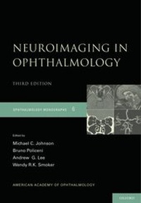
Neuroimaging in Ophthalmology
2. Auflage
Erschienen am 3. Februar 2011
Buch
Hardcover
128 Seiten
978-0-19-538161-0 (ISBN)
Beschreibung
Ophthalmologists are often the first clinicians to evaluate a patient harboring an underlying intraorbital or intracranial structural lesion. This unique position makes it particularly important for them to understand the basic mechanics, indications, and contraindications for the available orbital and neuroimaging studies (e.g., CT and MR imaging), as well as any special studies that may be necessary to fully evaluate the suspected pathology. It is equally important
for them to be able to communicate their imaging questions and provide relevant clinical information to the interpreting radiologist.
Since the publication of the original edition of this American Academy of Ophthalmology Monograph in 1992, new techniques and special sequences have improved our ability to detect pathology in the orbit and brain that are significant for the ophthalmologist. In this second edition of Monograph 6, Johnson, Policeni, Lee, and Smoker have updated the original content and summarized the recent neuroradiologic literature on the various modalities applicable to CT and MR imaging for ophthalmology.
They emphasize vascular imaging advances (e.g., MR angiography (MRA), CT angiography (CTA), MR venography (MRV), and CT venography (CTV) and specific MR sequences (e.g., fat suppression, fluid attenuation inversion recovery (FLAIR), gradient recall echo imaging (GRE), diffusion weighted imaging (DWI),
perfusion weighted imaging (PWI), and dynamic perfusion CT (PCT)). They have also included tables that outline the indications, best imaging recommendations for specific ophthalmic entities, and examples of specific radiographic pathology that illustrate the relevant entities.
The goal of this Monograph is to reinforce the critical importance of accurate, complete, and timely communication-from the prescribing ophthalmologist to the interpreting radiologist-of the clinical findings, differential diagnosis, and presumed topographical location of the suspected lesion in order for the radiologist to perform the optimal imaging study, and ultimately, to receive the best interpretation.
for them to be able to communicate their imaging questions and provide relevant clinical information to the interpreting radiologist.
Since the publication of the original edition of this American Academy of Ophthalmology Monograph in 1992, new techniques and special sequences have improved our ability to detect pathology in the orbit and brain that are significant for the ophthalmologist. In this second edition of Monograph 6, Johnson, Policeni, Lee, and Smoker have updated the original content and summarized the recent neuroradiologic literature on the various modalities applicable to CT and MR imaging for ophthalmology.
They emphasize vascular imaging advances (e.g., MR angiography (MRA), CT angiography (CTA), MR venography (MRV), and CT venography (CTV) and specific MR sequences (e.g., fat suppression, fluid attenuation inversion recovery (FLAIR), gradient recall echo imaging (GRE), diffusion weighted imaging (DWI),
perfusion weighted imaging (PWI), and dynamic perfusion CT (PCT)). They have also included tables that outline the indications, best imaging recommendations for specific ophthalmic entities, and examples of specific radiographic pathology that illustrate the relevant entities.
The goal of this Monograph is to reinforce the critical importance of accurate, complete, and timely communication-from the prescribing ophthalmologist to the interpreting radiologist-of the clinical findings, differential diagnosis, and presumed topographical location of the suspected lesion in order for the radiologist to perform the optimal imaging study, and ultimately, to receive the best interpretation.
Weitere Details
Reihe
Auflage
2nd Revised edition
Sprache
Englisch
Verlagsort
New York
USA
Zielgruppe
Für Beruf und Forschung
Ophthalmology, Radiographic, Radiology
Editions-Typ
Überarbeitete Ausgabe
Illustrationen
132 color halftone figures
Maße
Höhe: 260 mm
Breite: 181 mm
Dicke: 14 mm
Gewicht
492 gr
ISBN-13
978-0-19-538161-0 (9780195381610)
Copyright in bibliographic data and cover images is held by Nielsen Book Services Limited or by the publishers or by their respective licensors: all rights reserved.
Schweitzer Klassifikation
Weitere Ausgaben
Andere Ausgaben
Michael C. Johnson | Bruno Policeni | Andrew G. Lee
Neuroimaging in Ophthalmology
E-Book
12/2010
2. Auflage
OUP eBook
53,99 €
Als Download verfügbar
Michael C. Johnson | Bruno Policeni | Andrew G. Lee
Neuroimaging in Ophthalmology
E-Book
12/2010
2. Auflage
OUP eBook
53,99 €
Als Download verfügbar
Personen
Michael C. Johnson, M.D., F.R.C.S.C. is an Assistant Professor in the Department of Ophthalmology at the University of Alberta, Edmonton, Alberta, Canada.
Bruno Policeni, MD, Clinical Assistant Professor of Diagnostic Radiology-Neuroradiology, University of Iowa
Andrew G. Lee, M.D. is chair of the Department of Ophthalmology at The Methodist Hospital in Houston, Texas and is Professor of Ophthalmology in Neurology and Neurological Surgery at Weill Cornell Medical College. Dr Lee serves on the Editorial Board of 12 journals including the American Journal of Ophthalmology, the Canadian Journal of Ophthalmology, and Eye and he has published over 270 peer-reviewed articles, 40 book chapters, and three full textbooks in ophthalmology.
Wendy K. Smoker, MD, is a Professor of Radiology, Neurology, and Neurosurgery, and Division Director of Neuroradiology at the University of Iowa Hospitals and Clinics. Dr. Smoker has served on the Editorial Board of multiple Radiology journals, previously a
Deputy Editor of Radiology. She has authored or coauthored 144 scientific publications, presented 282 invited lectures, and authored or co-authored 47 book chapters.
Bruno Policeni, MD, Clinical Assistant Professor of Diagnostic Radiology-Neuroradiology, University of Iowa
Andrew G. Lee, M.D. is chair of the Department of Ophthalmology at The Methodist Hospital in Houston, Texas and is Professor of Ophthalmology in Neurology and Neurological Surgery at Weill Cornell Medical College. Dr Lee serves on the Editorial Board of 12 journals including the American Journal of Ophthalmology, the Canadian Journal of Ophthalmology, and Eye and he has published over 270 peer-reviewed articles, 40 book chapters, and three full textbooks in ophthalmology.
Wendy K. Smoker, MD, is a Professor of Radiology, Neurology, and Neurosurgery, and Division Director of Neuroradiology at the University of Iowa Hospitals and Clinics. Dr. Smoker has served on the Editorial Board of multiple Radiology journals, previously a
Deputy Editor of Radiology. She has authored or coauthored 144 scientific publications, presented 282 invited lectures, and authored or co-authored 47 book chapters.
Autor*in
Assistant Professor of OphthalmologyUniversity of Alberta
Clinical Assistant ProfessorUniversity of Iowa Hospitals and Clinics
Professor of Ophthalmology, Department of Ophthalmology, The Methodist Hospital, Houston, TX, University of Iowa Hospitals and Clinics, Department of Ophthalmology and Visual Sciences, University of Texas Medical Branch at Galveston
Professor of Radiology, Neurology, and NeurosurgeryUniversity of Iowa Hospitals and Clinics
Inhalt
Preface
Chapter 1 Magnetic Resonance Imaging
1-1 Physical Principles
1-2 Extraction of Spatial Information
1-3 T1 and T2 Defined
1-4 TR and TE Defined
1-5 T1 and T2 Relaxation
1-6 Factors Determining the Appearance of MRI Scans
1-7 T1-weighted Images
1-8 T2-weighted Images
1-9 CT versus MRI of Hemorrhage
1-10 Diffusion Weighted Imaging
1-11 Perfusion Weighted Imaging
1-12 High Resolution 3D Rapid Imaging
1-13 Paramagnetic Contrast Agents
1-14 Surface-Coil Techniques
1-15 Magnetic strength
1-16 Contraindications
Chapter 2 Computed Tomography
2-1 Physical Principles
2-2 Clinical Imaging Devices
2-3 Windows
2-4 Axial-Plane Imaging
2-5 Multiplanar Reconstruction
2-6 Computer Analysis
2-7 Contrast Enhancement in CT
2-8 Perfusion Computed Tomography
2-9 X-Ray Dosage
Chapter 3 Angiography and Other Specialized Imaging
3-1 MR and CT angiography and venography
3-2 MR spectroscopy
3-3 Functional MRI
Chapter 4 Ordering and Interpreting Images
4-1 Selection of Technique
4-2 Interpreting Images
4-3 Examination of Images
Summary
Suggested Readings
Chapter 1 Magnetic Resonance Imaging
1-1 Physical Principles
1-2 Extraction of Spatial Information
1-3 T1 and T2 Defined
1-4 TR and TE Defined
1-5 T1 and T2 Relaxation
1-6 Factors Determining the Appearance of MRI Scans
1-7 T1-weighted Images
1-8 T2-weighted Images
1-9 CT versus MRI of Hemorrhage
1-10 Diffusion Weighted Imaging
1-11 Perfusion Weighted Imaging
1-12 High Resolution 3D Rapid Imaging
1-13 Paramagnetic Contrast Agents
1-14 Surface-Coil Techniques
1-15 Magnetic strength
1-16 Contraindications
Chapter 2 Computed Tomography
2-1 Physical Principles
2-2 Clinical Imaging Devices
2-3 Windows
2-4 Axial-Plane Imaging
2-5 Multiplanar Reconstruction
2-6 Computer Analysis
2-7 Contrast Enhancement in CT
2-8 Perfusion Computed Tomography
2-9 X-Ray Dosage
Chapter 3 Angiography and Other Specialized Imaging
3-1 MR and CT angiography and venography
3-2 MR spectroscopy
3-3 Functional MRI
Chapter 4 Ordering and Interpreting Images
4-1 Selection of Technique
4-2 Interpreting Images
4-3 Examination of Images
Summary
Suggested Readings