

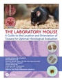
The Laboratory Mouse
A Guide to the Location and Orientation of Tissues for Optimal Histological Evaluation
1st Edition
Published on 21. March 2019
Book
Hardback
93 pages
978-0-367-17800-0 (ISBN)
Description
Key features:
High quality full color photographs and descriptive texts on the location and removal of the organs from the mouse
Instructive methods and clear visuals for trimming and orienting the organs for paraffin histology to obtain the best possible sections for analysis
Full color photomicrographs of the resulting section for each organ stained with hematoxylin and eosin demonstrating important features and landmarks for the histologist to ensure the optimal area for analysis is achieved
All in one, easy to use guide organized by individual organs of the laboratory mouse
Spiralbound for easy reference in the lab
This "one-stop" guide offers an essential resource for any academic, research or development operation where mouse necropsy and/or histology are performed. Connecting the reader 'from the mouse to the microscope', it provides a detailed guide for locating, trimming, orientating and embedding of the most frequently investigated tissues collected in the laboratory mouse. It shows where the organs reside in the mouse, how to trim and embed them as well as the resulting optimal sections. This guide brings together the wealth of scattered information into one high-quality text, the emphasis is on providing knowledge that will help histologists and scientists get better results in any downstream assays where ideal sections are needed.
More details
Language
English
Place of publication
London
United Kingdom
Publishing group
Taylor & Francis Ltd
Target group
Professional and scholarly
Professional Practice & Development
Illustrations
250 farbige Abbildungen
250 Illustrations, color
Dimensions
Height: 279 mm
Width: 216 mm
Weight
500 gr
ISBN-13
978-0-367-17800-0 (9780367178000)
Copyright in bibliographic data and cover images is held by Nielsen Book Services Limited or by the publishers or by their respective licensors: all rights reserved.
Schweitzer Classification
Other editions
Additional editions
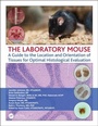
Jennifer Johnson | Brian DelGiudice | Dinesh Bangari
The Laboratory Mouse
A Guide to the Location and Orientation of Tissues for Optimal Histological Evaluation
Book
03/2019
1st Edition
CRC Press
€74.00
Shipment within 15-20 days

Jennifer Johnson | Brian DelGiudice | Dinesh Bangari
The Laboratory Mouse
A Guide to the Location and Orientation of Tissues for Optimal Histological Evaluation
E-Book
03/2019
1st Edition
CRC Press
€57.99
Available for download
Jennifer Johnson | Brian DelGiudice | Dinesh Bangari
The Laboratory Mouse
A Guide to the Location and Orientation of Tissues for Optimal Histological Evaluation
E-Book
03/2019
1st Edition
CRC Press
€57.99
Available for download
Persons
The author team are scientists at Global Discovery Pathology at Sanofi, USA. They have published in numerous journals and the lead author is a frequent speaker at the National Society for Histotechnology and other state societies.
Content
Introduction. Adrenal glands. Brain. Brain: Trimming for coronal sections. Brain: Trimming for sagittal sections. Diaphragm. Esophagus, Trachea and Thyroid. Eyes. Female: Ovaries. Female: Uterus (uterine horn), cervix, vagina. Femur. Heart. Kidneys. Liver and Gallbladder. Lung (inflated). Lymph nodes: Axillary. Lymph nodes: Mesenteric. Male: Epididymes. Male: Preputial gland. Male: Seminal vesicle, coagulating gland and prostate. Male: Testes. Pancreas. Pituitary gland. Quadriceps. Salivary glands. Sciatic nerve. Skin with (or without) mammary gland. Spinal cord. Spine. Spleen. Sternum. Stomach: Open method. Stomach: Whole method. Stifle joint. Thymus. Tongue. Urinary bladder. Intestines. Small intestine: Duodenum. Small intestine: Jejunum. Small intestine: Ileum. Large intestine: Cecum. Large intestine: Colon. Large intestine: Rectum. Materials and Methods. References.